Critically Appraised Topic (CAT): Back Pain and the Efficacy of the McKenzie Method of Mechanical Diagnosis and Therapy Management

Clinical Question: For patients with low back pain, is using the principle of Directional Preference through the McKenzie Method of Mechanical Diagnosis and Therapy Management efficacious?

Citation: Surkitt LD, Ford JJ, Hahne AJ, Pizzari T, McMeeken JM. Efficacy of directional preference management for low back pain: a systematic review. Phys Ther. 2012;92(5):652-665.

Clinical Bottom Line: Moderate evidence that directional preference management (DPM) was significantly more effective than a number of comparison treatments for pain, function, and work participation at short (12 months) follow-up. No trials found that DPM was significantly less effective than comparison treatment.

Summary of Key Evidence: The McKenzie Method is utilized in the treatment of LBP utilizing mechanical loading strategies (MLS). Directional Preference is the direction of MLS (exercise, etc.) that results in Centralization. Centralization is the proximal movement or abolition of distal symptoms in response to MLS. Directional Preference Management is individualized treatment based on the response to MLS.

  • Procedure: 6 RCT’s were selected from an initial 5932 references from 1950 thru January 2010 randomizing 474 participants. Subjects were male and female, greater than 17 years of age, with lumbar pain with or without leg pain or neurological signs with varying duration. The PEDro scale was used to determine the methodological quality and treatment effect, 95% confidence intervals were calculated and homogenous study data was pooled.
  • Outcome Measures: The Visual Analog Scale, numeric pain rating scale, Roland Morris Disability Questionnaire, Oswestry Disability Questionnaire and Functional Status Questionnaire were used with variability between the trials.
  • Secondary Outcome: The trials compared DPM with lumbar mobilization, manipulation, stabilization, strengthening, general conditioning exercises and advice to remain active. Different outcomes were reported for different time periods of follow-up.
  • Results: All trials except 1 were deemed high quality. Moderate evidence was found supporting the effectiveness of DPM when applied to participants with a Directional Preference in short and intermediate term follow ups. In high quality trials, DPM was shown significantly more effective than mid-range multidirectional exercises, advice and exercises in the opposite direction of DP.
  • Appraisal of Applicability, Internal validity, and Statistical Validity:
    • Threats-Low number of subjects, different trials reported different outcome measures, trials were heterogeneous so meta-analysis was not possible.
    • Strengths-DPM was compared to common clinical treatments with heterogeneous patient population.

Applicability to Patient: If a patient has a Directional Preference, or their pain improves with certain movements, they will benefit from a management approach utilizing these principles.

“My back hurts. Should I get an MRI?”

Back Pain in Durango CO | Tomsic Physical Therapy

This is a very common question I often get from my patients. They have come in for a Physical Therapy evaluation, and in the course fo the exam they ask about imaging.

Here are a few keys to keep in mind if you are a back pain sufferer and considering having imaging studies performed.

  1. An MRI, CT Scan or X-Ray cannot see pain. If you take a photograph of a person smiling, all that you can rightly say about that person is that they are smiling. You cannot accurately predict the exact emotion they are feeling at that time, or the thought that is going through their head. They could be thinking about all of the great skiing they are going to do this weekend, or maybe they are uncomfortable and smiling to hide their uneasiness. Diagnostic imaging is very much the same. “Pathologic” findings on imaging can be helpful guides in predicting what the pain generator COULD be. But it is impossible to say that a specific result is definitely, 100%, the cause of a patient’s pain. Skillful clinical examinations by a PT or physician can assist in providing an accurate diagnosis, but imaging alone is of questionable value.
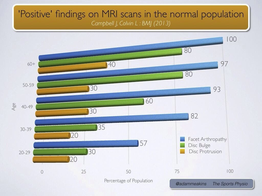
  2. Imaging can often be misleading. As I described in my first point, an image is simply an image. There is no way to “see” pain. The problem this causes is a high incidence of what are called “false positives.” False positives are any findings on imaging that could potentially be a problem, but are not the actual pain generators.In the image above, you can see the results of a study done by Jensen and colleagues that took MRI’s of non-painful participants. That’s right, these are people who DO NOT have back pain! In looking at the graph it is easy to see how a clinician could misdiagnose low back pain as a facet arthropathy or disc injury, when so many of these findings are also in normal pain free people. Similar studies have since been completed with corresponding results.

    Just because it is in an image, does not mean it is the source of pain.

    The scarier version of this problem comes when surgery is performed on a patient based on the findings in an imaging study, and after the long recovery process the patient’s original complaints remain. Unfortunately this does occur, though thankfully not frequently.

  3. Imaging can be costly. Low back pain has a yearly estimated health care cost of nearly $90 billion in the United States. This value does not include lost wages due to time off of work, or the personal expense back pain sufferers incur while trying over the counter remedies or tricks.The total cost is accumulated by prescriptions, imaging studies, surgeries, and rehabilitation. A 2003 study found that MRI’s performed early in a patent’s care, prior to PT evaluation, resulted in a 300% increase in surgical rates. And here is the kicker. Overall, there was no improvement in the patient’s condition even a year later. The patients in this study had to not only pay for an expensive MRI and then were subjected to costly, and more importantly, risky surgery. Only to be no better come a year later.

    It has been found that through judicious use of low back imaging, the total cost of an individuals care can be reduced by 53-60%, with quicker return to work and shorter recovery periods.

Now, keeping these things in mind, there are instances when imaging is necessary. The presence of progressive neurological symptoms such as changes in bowel or bladder function, or weakness is one major classification. Having a history of cancer with unexplained weight loss is another important one. Other collections of signs and symptoms exist that necessitate imaging studies to be performed, but a Physical Therapist is more than capable of screening for these, and is trained to do so during every initial examination.

So, when you begin to have an aching in your low back (as 80% of Americans will at one point in their life) be quick to get to your Physical Therapist. They will help you prevent unnecessary expense, rule imaging in or out based on your individual needs, and start you on a program to combat the problem right from the start.